Glaucoma (Types & Medication) Simplified for All - Doctor Solve
BUSINESS: Weekdays 6:00AM – 8:00PM PST
HOURSWeekends 7:00AM – 5:00PM PST
FIND US: Surrey, BC, Canada
#109–7938 128th Street V3W 4E8
CONTACT: +1-866-732-0305
[email protected]
  • No products in the cart.
Generic selectors
Exact matches only
Search in title
Search in content
Search in posts
Search in pages

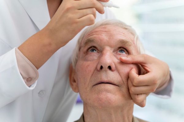
Glaucoma Medications

What is Glaucoma?

A glaucoma is a group of eye diseases that gradually steals sight without warning and often without symptoms. Vision loss is caused by damage to the optic nerve. This nerve acts like an electric cable with over a million wires and is responsible for carrying the images we see to the brain.

It was once thought that high intraocular pressure (IOP) was the main cause of this optic nerve damage. Although IOP is a risk factor, we now know that other factors must also be involved because even people with “normal” IOP can experience vision loss from glaucoma.

Different types of Glaucoma

The two main types of glaucoma are open-angle glaucoma, or primary open-angle glaucoma (POAG), and angle closure glaucoma.

Primary Open Angle Glaucoma

This is the most common form of glaucoma, affecting about three million Americans. It happens when the eye’s drainage canals become clogged over time.

The inner eye pressure (also called intraocular pressure or IOP) rises because the correct amount of fluid can not drains out of the eye. With open-angle glaucoma, the entrances to the drainage canals are clear and should be working correctly. The clogging problem occurs inside the drainage canals, like the clogging that can occur inside the pipe below the drain in a sink.

Most people have no symptoms and no early warning signs. If open-angle glaucoma is not diagnosed and treated, it can cause a gradual loss of vision. This type of glaucoma develops slowly and sometimes without noticeable sight loss for many years. It usually responds well to medication, especially if caught early and treated.

Angle Closure Glaucoma

This type of glaucoma is also known as acute glaucoma or narrow-angle glaucoma. It is much rarer and is very different from open-angle glaucoma in that the eye pressure usually goes up very fast. This happens when the drainage canals get blocked or covered over, like the clog in a sink when something is covering the drain. With angle closure glaucoma, the iris and cornea are not as wide and open as they should be. The outer edge of the iris bunches up over the drainage canals when the pupil enlarges too much or too quickly. This can happen when entering a dark room.

A simple test can be used to see if your angle is normal and wide or abnormal and narrow. Treatment of angle closure glaucoma usually involves surgery to remove a small portion of the outer edge of the iris. This helps unblock the drainage canals so that the extra fluid can drain.

Usually, surgery is successful and long-lasting. However, you should still receive regular check-ups. Symptoms of angle-closure glaucoma may include headaches, eye pain, nausea, rainbows around lights at night, and very blurred vision.

Secondary Glaucoma

Glaucoma can occur as the result of an eye injury, eye inflammation, tumor, or in advanced cases of cataracts or diabetes. It can also be caused by certain drugs such as steroids. This form of glaucoma may be mild or severe. The type of treatment will depend on whether it is the open angle or angle closure glaucoma.

Normal-Tension Glaucoma (NTG)

Normal tension glaucoma is also known as low tension glaucoma or normal pressure glaucoma. In this type of glaucoma, the optic nerve is damaged even though intraocular pressure (IOP) is not very high. Doctors do not know why some people’s optic nerves are damaged even though they have what is considered to be “normal” (between 12-22 mm Hg) pressure levels.

Those at higher risk for this form of glaucoma are people with a family history of normal tension glaucoma, people of Japanese ancestry, and people with a history of systemic heart disease, such as irregular heart rhythm. Normal tension glaucoma is usually detected after an examination of the optic nerve.

Pigmentary Glaucoma

This is a form of secondary open-angle glaucoma. It occurs when the pigment granules in the back of the iris (the colored part of the eye) break into the clear fluid produced inside the eye. These tiny pigment granules flow toward the drainage canals in the eye and slowly clog them, causing eye pressure to rise. Treatment usually includes medications or surgery.

Cataracts and Glaucoma

Both cataracts and glaucoma can be a natural part of the aging process. Many people over 60 may have both. Otherwise, the two are not associated. Except for glaucoma due to secondary causes such as trauma or steroids, glaucoma does not cause cataracts and cataracts do not cause glaucoma. While glaucoma is most often a problem with drainage, a cataract is a clouding of the eye’s lens allowing less light to pass through.

Treating Glaucoma

Regular glaucoma check-ups include two routine eye tests: tonometry and ophthalmoscopy.

Tonometry

The tonometry test measures the inner pressure of the eye. Usually, drops are used to numb the eye. Then the doctor or technician will use a special device that measures the eye’s pressure.

Ophthalmoscopy

Ophthalmoscopy is used to examine the inside of the eye, especially the optic nerve. In a darkened room, the doctor will magnify your eye by using an ophthalmoscope (an instrument with a small light on the end). This helps the doctor look at the shape and color of the optic nerve.

If the pressure in the eye is not in the normal range, or if the optic nerve looks unusual, then one or two special glaucoma tests will be done. These two tests are called perimetry and gonioscopy.

Perimetry

The perimetry test is also called a visual field test. During this test, you will be asked to look straight ahead and then indicate when a moving light passes your peripheral (or side) vision. This helps draw a “map” of your vision.

Gonioscopy

Gonioscopy is a painless eye test that checks if the angle where the iris meets the cornea is open or closed, showing if either open-angle or closed-angle glaucoma is present.

It is important to have your eyes examined regularly. Your eyes should be tested at:

  • Ages 35 and 40
  • After age 40, every two to four years
  • After age 60, every one to two years
  • Those with any high-risk factors, every one to two years after age 35

Medications for Glaucoma

In these tables ‘brand name’ denotes the manufacturer’s name for the medication, and ‘generic’, is the generic name.

Adrenergic Type Drugs

Company:
Brand Name:
Generic:
Allergan Inc.
Propine
Dipivefin HCI Ophthalmic Solution USP, 0.1%
Increases drainage of intraocular fluid

Side effects Allergic reactions to these drops are frequent. Blurred vision can also occur. Vision can also be decreased in people who have had cataracts removed. Headaches and burning of the eyes are also common. Cardiac side effects may include rapid heart rate or fluctuations in heart rhythm.

Alpha Agonist

Company:
Brand Name:
Generic:
Alcon Laboratories, Inc.
Iopidine
Apraclonidine HCI 0.5%, 1%

Company:
Brand Name:
Generic:
Allergan Inc.
Alphagan
Brimonide tartrate 0.15%, 0.2%

Works to both decrease production of fluid and increase drainage Side effects can include burning or stinging upon instillation of the eye drop, fatigue, headache, drowsiness, dry mouth, and dry nose.

Beta Blockers

Company:
Brand Name:
Generic:
Akorn Ophthalmics
Timolol Maleate USP
Timolol Maleate 0.5%

Company:
Brand Name:
Generic:
Alcon Laboratories Inc.
Betoptic S
Betaxolol HCI 0.25%, 0.5%

Company:
Brand Name:
Generic:
Allergan Inc.
Betagan
Levobunolol HCI ophthalmic solution, USP 0.25%, 0.5%

Company:
Brand Name:
Generic:
Bausch & Lomb Pharmaceuticals
OptiPranolol
Metipranolol 0.3%

Company:
Brand Name:
Generic:
Johnson & Johnson
Betimol
Timolol hemihydrate 0.25%, 0.5%

Company:
Brand Name:
Generic:
Merck & Co. Inc.
Timoptic-XE
Timolol maleate ophthalmic gel forming solution 0.25%, 0.5%

These medications decrease the production of intraocular fluid.

Side effects can include low blood pressure, reduced pulse rate, and fatigue. Beta-blockers can also cause shortness of breath in people who have a history of asthma or other respiratory disorders. Additionally, beta blockers can change cardiac activity by decreasing the amount of blood the heart pumps out, which may reduce the pulse rate and/or slow down the hearts response rate during exercise. Rare side effects include reduced libido and depression.

Carbonic Anhydrase Inhibitors

Company:
Brand Name:
Generic:
Alcon Laboratories Inc.
Azopt
Brinzolamide ophthalmic suspension 1%

Company:
Brand Name:
Generic:
Merck & Co. Inc.
Trusopt
Dorzolamide HCI 2%

Company:
Brand Name:
Generic: Brand Name:
Generic:
Wyeth Ayerst
Diamox Sequels
Acetazolamide
Neptazane
Methazolamide

This medication decreases the production of intraocular fluid.

Side effects of the pill form of these medications can include tingling or loss of strength in the hands and feet, upset stomach, mental fuzziness, memory problems, depression, kidney stones, and frequent urination. Since the eyedrop form of this medication is relatively new, long-term studies are yet to be completed. Current effects of the eyedrop include stinging, burning, and another eye discomfort.

Cholinergic (Miotic)

Company:
Brand Name:
Generic:
Brand Name:
Generic:
Brand Name:
Generic:
Alcon Laboratories, Inc.
Isopto Carpine
Pilocarpine HCI 1%, 2%, 4%, 6%, 8%
Isopto Carbachol
Carbachol 0.75%, 1.5%, 3%
Pilopine HS Gel
Pilocarpine HCI gel 4%

Company:
Brand Name:
Generic:
Bausch & Lomb Pharmaceuticals
Pilocarpine HCI ophthalmic solution USP
Pilocarpine HCI ophthalmic solution USP ) 0.5%, 1%, 2%, 3%, 4%, 6%

These increase the drainage of intraocular fluid.

Side effects Many people who use these medications complain of dim vision, especially at night or in darkened areas such as movie theaters. This is due to constriction of the pupil. Miotics increase the drainage of intraocular fluid by making the pupil size smaller, thereby increasing the flow of intraocular fluid from the eye.

Cholinesterase Inhibitor

Company:
Brand Name:
Generic:
Wyeth Ayerst
Phospholine Iodide
Echothiophate

Increases drainage of intraocular fluid.
Side effects same as cholinergic.

Combined
Company:
Brand Name:
Generic:
Merck & Co. Inc
Cosopt
Dorzolomide HCI timolol maleate

Increases drainage of intraocular fluid

Side effects Allergic reactions to these drops are frequent. Blurred vision can also occur. Vision can also be decreased in people who have had cataracts removed. Headaches and burning of the eyes are also common. Cardiac side effects may include rapid heart rate or fluctuations in heart rhythm.

Prostaglandin Analogs

Company:
Brand Name:
Generic:
Alcon Laboratories, Inc.
Travatan
Travaporst 0.0004%

Company:
Brand Name:
Generic:
Allergan, Inc.
Lumigan
Bimatoprost 0.03%

Company:
Brand Name:
Generic:
Pfizer, Inc.
Xalatan
Latanoprost 0.0005%

Decreases production of intraocular fluid.

Side effects In initial studies, between 5% and 15% of people who used this medication reported a gradual change in eye color, due to an increased amount of brown pigment in the iris of the treated eye. The change in eye color occurs slowly and may not be noticeable for several months to years. Other side effects can include stinging, blurred vision, eye redness, itching, and burning. These medications are new to the market and long-term follow-up of people who use them is not yet available.

Surgery

Laser

What is laser surgery? The type of laser surgery used depends on the type of glaucoma and the general health of the eye. Laser surgeries have become important in the treatment of different eye problems and medical conditions.

During the laser surgery, the eye is numbed so that there is little or no pain. The eye doctor then holds a special lens to the eye. The laser beam is aimed into the eye, and a bright light – like a camera flash – is seen.

Are there risks associated with glaucoma laser surgeries?

Laser surgery is still surgery, and can carry some risks. Some people experience a short-term increase in their intraocular pressure (IOP) soon after surgery. In others who require YAG CP (Cyclophoto-Coagulation) surgery, there is a risk of the IOP dropping too low to maintain the eye’s normal metabolism and shape. But the use of anti-glaucoma medication before and after surgery can help to reduce this risk.

The most common glaucoma laser surgeries are:

Laser Peripheral Iridotomy (LPI):

Often used in people with narrow-angle glaucoma. Narrow-angle glaucoma occurs when the angle between the iris and the cornea in the eye is too small. This causes the iris to block fluid drainage, which makes the inner eye pressure increase. LPI makes a small hole in the iris, allowing it to fall back from the fluid channel and helping the fluid drain.

Argon Laser Trabeculoplasty (ALT):

Used in people with primary open-angle glaucoma (POAG). The laser beam opens the fluid channels of the eye, helping the drainage system to work better. In many cases, medication will still be needed.

It has successfully lowered eye pressure in up to 75% of patients treated.

Selective Laser Trabeculoplasty (SLT):

Used in people with primary open-angle glaucoma (POAG). A type of laser surgery that uses a combination of frequencies allowing the laser to work at very low levels. It treats specific cells “selectively” and leaves untreated portions of the trabecular meshwork intact. For this reason, it is believed that SLT, unlike other types of laser surgery, may be safely repeated many times.

Nd:YAG Laser Cyclophoto-coagulation (YAG CP):

Used in people with severe glaucoma damage that is not being managed by standard glaucoma surgery. This surgery destroys part of the ciliary body, the part of the eye that produces intraocular fluid.

FAQs on laser surgery

Is there pain or discomfort with glaucoma laser surgery?

There is a slight stinging sensation associated with LPI and ALT. In YAG CP laser surgery, a local anesthetic is used to numb the eye. Once the eye has been numbed, there should be little or no pain and discomfort.

What are the long-term benefits of having glaucoma laser surgery? Will a repeat procedure be needed?

Glaucoma laser surgeries help to lower the IOP in the eye. The length of time the IOP is lowered depends on the type of laser surgery, the type of glaucoma, age, race, and many other factors. Some people may need the surgery repeated to get better control of the IOP, depending on the type of laser surgery done.

Is it common to have laser surgery over two sessions?

For people requiring ALT surgery, half of the fluid channels are usually treated first. Depending on the person’s eye, the other fluid channels can be treated at different times. This method prevents over-correction and lowers the risk of an increased IOP after the surgery.

Will laser surgery eliminate the need for medications?

In most cases, medications are still necessary to control and maintain eye pressure. However, surgery may lessen the amount of medication needed.

How much recovery time is needed after laser surgery?

Laser surgery is usually performed in an eye doctor’s office or eye clinic. Before the surgery, the eye will be numbed with medicine. The eye may be a bit irritated and vision may be slightly blurry after laser surgery. A ride home on the day of the surgery should be arranged.

In general, patients can resume their normal daily activities the day after the surgery. But it is important to discuss any questions or concerns about laser surgery with the eye doctor.

Does glaucoma laser surgery increase the risk of developing cataracts?

There is a small risk of developing cataracts after some types of laser surgery for glaucoma. However, the potential benefits of the surgery usually outweigh any risks.

There is a common myth that lasers can be used to remove cataracts; this is not the case except in experimental studies. After a cataract has been taken out with conventional cutting surgery, there often remains an outer membrane lens capsule. This membrane can slowly thicken and cloud vision, just as the cataract did. Laser surgery can open this membrane, helping to clear vision without an operation. This laser procedure is called a capsulotomy.

Filtering surgery

What is filtering microsurgery?

When medicines and laser surgeries do not lower eye pressure adequately, therefore doctors may recommend a procedure called filtering microsurgery (sometimes called conventional or cutting surgery).

In filtering microsurgery, a tiny drainage hole is made in the sclera (the white part of the eye) in a procedure called a trabeculectomy or a sclerostomy. The new drainage hole allows fluid to flow out of the eye and helps lower eye pressure. This prevents or reduces damage to the optic nerve.

Is there pain during the microsurgery?

In most cases, there is no pain involved. The surgery is usually done with a local anesthetic and relaxing medications. Often a limited type of anesthesia, called intravenous (I.V.) sedation, is used.

In addition, an injection is given around or behind the eye to prevent eye movement. This injection is not painful when I.V. sedation is used first. The patient will be relaxed and drowsy and will not experience any pain during surgery.

How does the eye doctor determine if filtering surgery is needed?

Often, laser surgery is recommended before filtering microsurgery, unless the eye pressure is very high or the optic nerve is badly damaged. During laser surgery, a tiny but powerful beam of light is used to make several small scars in the eye’s trabecular meshwork (the eye’s drainage system). The scars help increase the flow of fluid out of the eye.

In contrast, filtering microsurgery involves creating a drainage hole with the use of a small surgical tool. When laser surgery does not successfully lower eye pressure, or the lowering effects wear off, the doctor may recommend filtering microsurgery.

What is the success rate?

Most of the related studies document follow-up for one year. In those reports, it shows that in older patients, glaucoma filtering surgery is successful in about 70-90% of cases, for at least one year.

Occasionally, the surgically-created drainage hole begins to close and the pressure rises again. This happens because the body tries to heal the new opening in the eye as if the opening were an injury. This rapid healing occurs most often in younger people because they have a stronger healing system. Anti-wound healing drugs, such as mitomycin-C and 5-FU, help slow down the healing of the opening. If needed, glaucoma filtering surgery can be done several times in the same eye.

Is this an outpatient procedure or is an overnight stay in the hospital needed?

Usually, filtering surgery is an outpatient procedure, requiring no overnight hospital stay. Within a few days after surgery, the eye doctor will need to check on the eye pressure. The doctor will also look for any signs of infection or an increase in inflammation.

How long is the recovery time?

For at least one week after surgery, patients are advised to keep water out of the eye. Most daily activities can be done, however, it is important to avoid driving, reading, bending, and doing any heavy lifting. Since each case is different, patients should check with their doctor for specific advice.

How will the eye look after surgery? Is there a noticeable scar or redness?

The eye will be red and irritated shortly after surgery, and there may be increased eye tearing or watering. The inner eye fluid flows through the surgically-created hole and forms a small blister-like bump called a bleb. The bleb, usually located on the upper surface of the eye, is covered by the eyelid and is usually not visible.

Will vision be changed? Will glaucoma medications still need to be taken?

There may be some vision changes, such as blurred vision, for about six weeks after the surgery. After that time, vision will usually return to the same level it was before surgery.

Vision can sometimes improve after surgery in people who were previously using pilocarpine. When pilocarpine drops are no longer taken, the pupil returns to normal size, allowing more light to enter the eye.

In a few cases, the vision may be worse due to very low pressure. Cataracts or wrinkles in the macula area of the eye may develop.
In terms of vision correction, a patient’s glasses or contact lenses may need to be changed after surgery. Both gases permeable and soft contact lenses may be worn after glaucoma filtering surgery. There may be fitting problems due to the bleb. Special care is needed to avoid infection of the bleb. Contact lens users should discuss these problems with their eye doctor following surgery.
Doctorsolve Healthcare Solution site strives to provide you with timely, accurate information, which is not intended for diagnosis or treatment.

Unfortunately, due to a power outage some of the features available on Doctorsolve may not be online. We know how important it is to have affordable prescription medication and we’re working as fast as we can to restore our call center, email, and online chat services.